Pituitary Cystic Lesion Mri
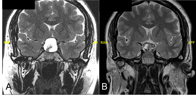
MRI T2-weighted axial image a T1-weighted post-gadolinium axial b and coronal images c demonstrate an extra-axial expansive lesion dotted circles mildly hyperintense with heterogenous contrast enhancement surrounded by cystic components. The presence of a fluid-fluid level a hypointense rim on T2-weighted images septation and an off-midline location were more common with pituitary adenomas whereas the presence of an intracystic nodule was more common with Rathke cleft cysts.

Preoperative Postcontrast Pituitary Mri Showing An Intrasuprasellar Download Scientific Diagram
She was taken to the emergency room where an MRI was performed and demonstrated a left orbital lesion extending into the superior orbital fissure.

Pituitary cystic lesion mri. Calvarial lesions are radiologically evaluated with CT and MRI. My MRI was loaded with white matter lesions neurologist said he was frightened for the number and size of of them and I was diagnosed with probable MS. Tumor lesions of the pituitary gland and tissues surrounding the Turkish saddle. The mass is a histologically proven pituitary macroadenoma which presented initially with a large cystic subfrontal extension that was successfully resected in April of 2006. Occasionally headaches or pituitary dysfunction may occur. Cystic hygromas are typically noticed and diagnosed before two years old presenting as soft.
Cystic fibrosis CF. The Journal of Pediatrics is an international peer-reviewed journal that advances pediatric research and serves as a practical guide for pediatricians who manage health and diagnose and treat disorders in infants children and adolescentsThe Journal publishes original work based on standards of excellence and expert review. An adenoma is a benign tumor of epithelial tissue with glandular origin glandular characteristics or both. Magnetic Resonance Imaging MRI a special imaging technique that uses a powerful magnet and a computer to provide clear images of soft tissues. To promote equity and diversity among authors reviewers and editors. However it is often adenoma vs.
Incidental adrenal masses on imaging are common 06 to 13 of all abdominal CT. To provide a platform for discussion of current ideas in urologic education patient engagement. Magnetic resonance imaging using contrast can detect many abnormalities in the brain. After redoing the MRI with contrast the lesions were seen again and so was a pituitary tumor. Accurate coding information must be provided with claims to differentiate CT andor MRI scans from other radiology services and to make coverage determinations. Further testing was requested to verify definite MS.
After at least 12 hours MRI is superior to CT in detecting haemorrhage. But for sub-acute and chronic stages of pituitary apoplexy brain MRI is considerably better than CT. Pituitary gland a small oval gland located at the base of the brain that. Tissues that are well-visualized using MRI include the brain and spinal cord abdomen and joints. The main indications for diagnostic manipulation are. A review of the ophthalmological features.
On T2-weighted images optic-nerve glioma is hyperintense to cerebral cortex and may appear heterogeneous secondary to cystic degeneration. They can be found anywhere on the body but classically presents in the axilla or posterior triangle of the neck. 67-year-old man presented to the strabismus clinic with increased difficulty reading for the past year. The hypothalamus from the Greek hypo meaning below and thalamus meaning bed is that part of the diencephalon located below the thalamusIt is a small but highly complex structure in the brain that controls many important body functions 1 4Magnetic resonance MR imaging is the modality of choice in evaluating the hypothalamic. MRI may show cystic degeneration if present. Sufficient documentation such as history and physical notes laboratory results signs and symptoms of the disease to warrant the diagnostic test and to support the claim of reasonable.
It offers diagnostic and imaging services such as ultrasound x. Tumors of the cerebellopontine angle. In patients with congenital growth hormone deficiency an MRI will help to identify abnormal pituitary anatomy rather than for definitive diagnosis. Empty sella syndrome may occur as a primary disorder for which the cause is unknown idiopathic or as a secondary disorder in which it occurs due to an underlying condition or disorder such as a pituitary tumor or trauma in the pituitary region. Gliomas enhance variably and a complete lack of enhancement can also occur. In the article Bone Tumors - Differential diagnosis we discussed a systematic approach to the differential diagnosis of bone tumors and tumor-like lesions.
This tumor was ignored. This patient has been observed closely for 25 years and despite obvious mass effect he has no visual complaints and the neuro-ophthalmologic evaluation is normal. CT is the most accurate method for evaluating bone destruction of the inner and outer tables the lytic or sclerotic nature of the lesion and for the evaluation of mineralised tumour matrix 123 6MRI is best to depict marrow involvement of the diploe and to evaluate the associated soft tissue. Incidence of pulmonary embolism and impact on mortality in patients with malignant melanoma. The Journal seeks to publish high. Ganesh Diagnostic is a renowned diagnostic centres and pathology labs Rohini in the state of Delhi in India.
Magnetic resonance imaging MRI is usually less efficient than CT in the acute stage of pituitary apoplexy. Magnetic resonance imaging MRI with pituitary protocol is the preferred study to characterize lesions even if computed tomography CT. MRI is useful in showing intracranial extension. It can view organs that may be obscured by bone or foreign bodies on conventional x-rays or CT scans. In this article which is the first in a series of three we will discuss the most common bone tumors and tumor-like lesions in alphabethic order. With special sequence MRI is useful for early detection.
Adenomas can grow from many glandular organs including the adrenal glands pituitary gland thyroid prostate and othersSome adenomas grow from epithelial tissue in nonglandular areas but express glandular tissue structure as can happen in familial polyposis coli. Tumors and metastases of the brain. It is capable of showing the tissues from multiple viewpoints and is. Some of these lesions are easily identified by radiographic appearance. According to their biological behavior lesions of the adrenal grands can be classified into being benign or being malignant including primary or metastatic Table 1Different lesions have different treatment options and clinical prognoses so it is of great clinical value to make a differential diagnosis based on computed tomography CT and. The mass is located along the course of the right hypoglossal nerve.
Multiple logistic regression analysis showed that cystic pituitary adenomas and Rathke cleft. MRI is superior in most cases in which differentiation of soft tissues is necessary. A cystic hygroma also known as a cystic lymphangioma is a benign fluid-filled sac caused by a malformation of the lymphatic system. Quantitative parameters of magnetic resonance imaging cannot predict human epidermal growth factor receptor 2 HER2 status in rectal cancer. An MRI is usually completed to assess for tumors and a bone density test will provide information about bone strength and possible disorders eg osteopenia osteoporosis. Differential diagnosis include adenoma myelolipoma cyst lipoma pheochromocytoma adrenal cancer metastatic cancer hyperplasia and tuberculosis.
The mission of Urology the Gold Journal is to provide practical timely and relevant clinical and scientific information to physicians and researchers practicing the art of urology worldwide.
Fig 5 Differentiation Between Cystic Pituitary Adenomas And Rathke Cleft Cysts A Diagnostic Model Using Mri American Journal Of Neuroradiology

Mri Pituitary With Contrast Revealing A Cystic Lesion In The Pituitary Download Scientific Diagram
Fig 6 Differentiation Between Cystic Pituitary Adenomas And Rathke Cleft Cysts A Diagnostic Model Using Mri American Journal Of Neuroradiology
Pituitary Cysts In Childhood Evaluated By Mr Imaging American Journal Of Neuroradiology

Post Contrast Mri Revealed A A Cystic Pituitary Adenoma Sagittal Download Scientific Diagram
Rathke S Cleft Cyst Pietro Mortini

Cystic Pituitary Macroadenoma Rathke S Cleft Cyst With Intracystic Nodule Springerlink
Differentiation Between Cystic Pituitary Adenomas And Rathke Cleft Cysts A Diagnostic Model Using Mri American Journal Of Neuroradiology

Cystic Pituitary Macroadenoma Rathke S Cleft Cyst With Intracystic Nodule Springerlink
Hemorrhagic Pituitary Adenoma Versus Rathke Cleft Cyst A Frequent Dilemma Ajnr Blog

Magnetic Resonance Imaging Mri Images Of A Cystic Sellar Lesion Found Download Scientific Diagram
T2 Hypointense Signal Of Rathke Cleft Cyst American Journal Of Neuroradiology

Rathke Cleft Cyst Presenting As Recurrent Pituitary Apoplexy An Unusual Case Presentation And Clinical Course
Differentiation Between Cystic Pituitary Adenomas And Rathke Cleft Cysts A Diagnostic Model Using Mri American Journal Of Neuroradiology
Posting Komentar untuk "Pituitary Cystic Lesion Mri"