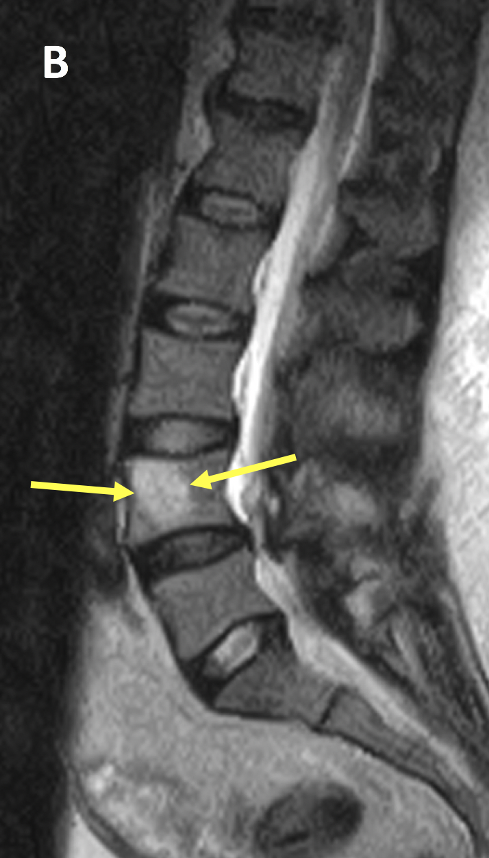
Small Hemangioma In The L1 Vertebral Body
Vertebral hemangiomas are common tumors incidentally found on neuro-imaging mostly in the thoracic region. A spinal hemangioma or a hemangioma in spine is a benign tumor that may develop in the bony segments of the spinal column.
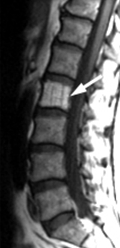
Vertebral Hemangioma Radsource
Hemangioma is a benign tumor that occurs in the endothelial lining of the blood vessels.

Small hemangioma in the l1 vertebral body. Hemangioma Vertebral When symptomatic they can cause pain and myelopathy by intra-spinal bleeding bony expansion or extra-osseous extension into surrounding soft tissue or the posterior neural elements. These growths classically appear in the thoracic and lumbar spine located in the mid to lower back. Roentgenograms usually show multiple or multilocular lytic lesions. It can occur at all ages but is most common in the fourth and fifth decades of life and has a female preponderance 31 1. The term hemangioma refers to a mass of blood vessels that commonly occur on the subcutaneous tissues. Of a vertebral hemangioma can be difficult.
Despite being the most common tumor of the spine vertebral hemangioma is rarely symptomatic in adults. Unless it is causing you pain doubtful it would be just forget about it- its benign. They typically have a benign asymptomatic course but rarely they will enlarge and can cause pain fracture etc. D1809 is a billablespecific ICD-10-CM code that can be used to indicate a diagnosis for reimbursement purposes. Spinal hemangiomas usually appear in the middle of your back thoracic area or your lower back lumbar area. The disorder is usually localized in the thoracic th12 and lumbar l1-l4 spine affecting one or several vertebrae.
Given below is some information on what causes an atypical hemangioma in spine and how it can be treated. Learn More About The Continuous Innovation Happening in the Oncology Community. Hemangiomas are noncancerous benign tumors made of abnormal blood vessels. A lumbar hemangioma is a benign blood vessel tumor that grows along one or more vertebra of the lower back. And should i be concerned Answered by Dr. All neoplasms are classified in this chapter.
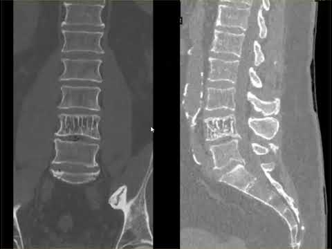
In actuality hemangiomas are common lesions with approximately 10 of autopsy cases having vertebral hemangiomas in one study. Lesions generally become symptomatic when there is neural arch expansion vertebral body enlargement or direct compression of the thecal sac or nerve roots 7812 and greater than 50 of patients with symptomatic vertebral hemangiomas present with pain 13. This is the American ICD-10-CM version of D1809 - other international versions of ICD-10 D1809 may differ. A spinal hemangioma refers to a tumor of the spine. What does this mean. The classic CT appearance of a hemangioma is a well-circumscribed lesion with a sharp transition zone and sclerotic margin within the vertebral body.
Spinal hemangiomas are the most common primary tumor of the spine1 Usually benign this lesion is of vascular origin and like hemangiomas in other parts of the body usually involves a proliferation of normal capillary and venous structures2 According to one study they have been identified in about 11 of patients at general autopsy3. 1 cm is small. A spinal hemangioma is a benign vascular tumor of the spine. Bone hemangiomas are benign malformed vascular lesions overall constituting fewer than 1 of all primary bone neoplasms. A spinal hemangioma is a tumor that mostly occurs in the thoracic and the lumbar spine. While the tumor is not dangerous it can cause pain and discomfort and treatment may be recommended for these reasons.
They say it is likely benign. Hemangiomas are largely asymptomatic and thus most are never discovered leading some to describe these lesions as rare. The 2022 edition of ICD-10-CM D1809 became effective on October 1 2021. Most hemangiomas involve the vertebral body or skull and involvement of other bones is rare. Causes and types of hemangiomas. However involvement of long and short tubular bones has been reported.
These lesions typically do not require treatment. Usually there is damage to the vertebral bodies but the possibility of tumor growth and cartilage layers. Hemangioma of the l1 vertebral body. Vertebral hemangiomas are observed throughout any age although most are diagnosed in people within their 50s alongside a higher presence in females with a 115 male-to-female ratio. Intraosseous hemangiomas are usually found in the vertebral column and rarely seen in the calvarium. The peak incidence is in the fifth decade.
Hemangioma is a vascular tumor which is a tangle of interwoven and modified vessels. Hemangioma is the most common benign vascular tumor of bone. Your skin muscles internal organs or bones. Theyre common and can occur anywhere in the body. This is why most people do not realize that they have this condition unless an imaging test for another condition reveals its presence. Ad Find Research Into the Innovative Future of Cancer Therapy For Patients with Solid Tumors.
Approximately 50 of osseous hemangiomas are found in the. Intraosseous hemangioma is a rare bone tumor accounting for 07 to 10 of all bone tumors. Also referred to as vertebral hemangioma it is non-cancerous and has few symptoms. These are birthmarks on the bone instead of the skin. They often present in the vertebral body of the thoracic and lumbar spine with potential to extend into the posterior arch. This type of tumor is most frequently diagnosed in patients between the ages of 30 and 50 and may not cause noticeable symptoms according to Scoliosis and Spine Associates.
Most bone hemangiomas are on the spine and develop after age 50.
Spine Lecture Hemangioma Of Spine Youtube
Vertebral Hemangioma Radsource
Vertebral Hemangioma Radsource
Differentiating Atypical Hemangiomas And Metastatic Vertebral Lesions The Role Of T1 Weighted Dynamic Contrast Enhanced Mri American Journal Of Neuroradiology

Vertebral Hemangioma Radsource
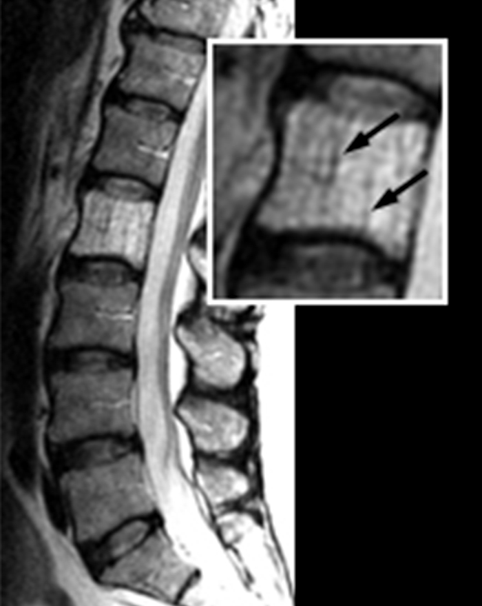
Vertebral Hemangioma Radsource
Hemangioma Vertebral Mri Online
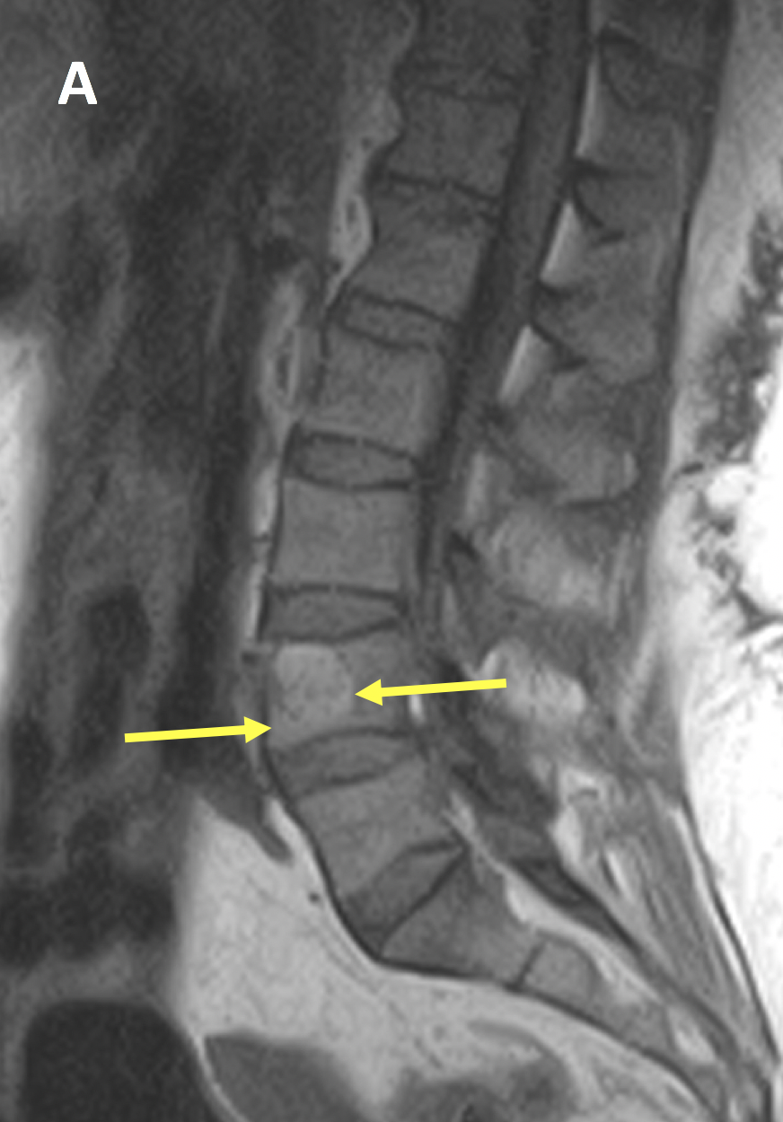
Aggressive Vertebral Hemangioma Involving L1 Vertebra In A 54 Year Old Download Scientific Diagram

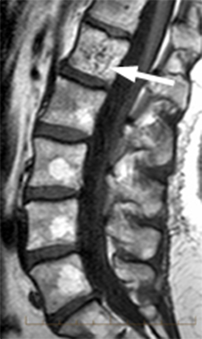
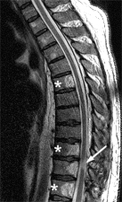
Atypical Mr Imaging Features Of Vertebral Hemangioma Involving The T12 Download Scientific Diagram

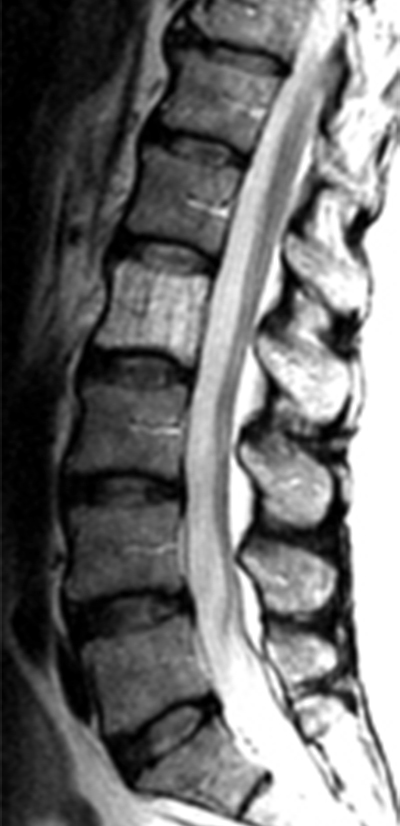
Typical Mr Imaging Features Of Vertebral Hemangiomas A Sagittal Download Scientific Diagram

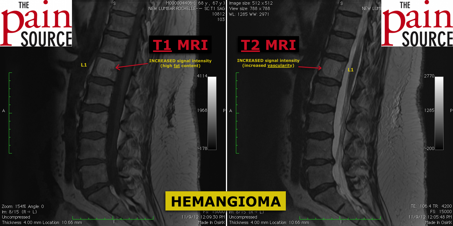
Hemangioma In The Spine The Pain Source Makes Learning About Pain Painless
Hemangioma Vertebral Mri Online
Long Term Outcome Of Treatment Of Vertebral Body Hemangiomas With Direct Ethanol Injection And Short Segment Stabilization The Spine Journal

Vertebral Hemangioma Wikipedia


Posting Komentar untuk "Small Hemangioma In The L1 Vertebral Body"