Does A Lumbar Spine Mri Show Kidneys
MRI and x-ray for low back pain are surprisingly unreliable1 because things like bulging discs usually arent a big deal2 most back pain goes away on its own3 and trigger points muscle knots are common and can be worrisomely intense but arent dangerous4 Most patients are much better off when they feel confident about these things. MRI magnetic resonance imaging is a noninvasive diagnostic test that takes detailed images of the soft tissues of the body.
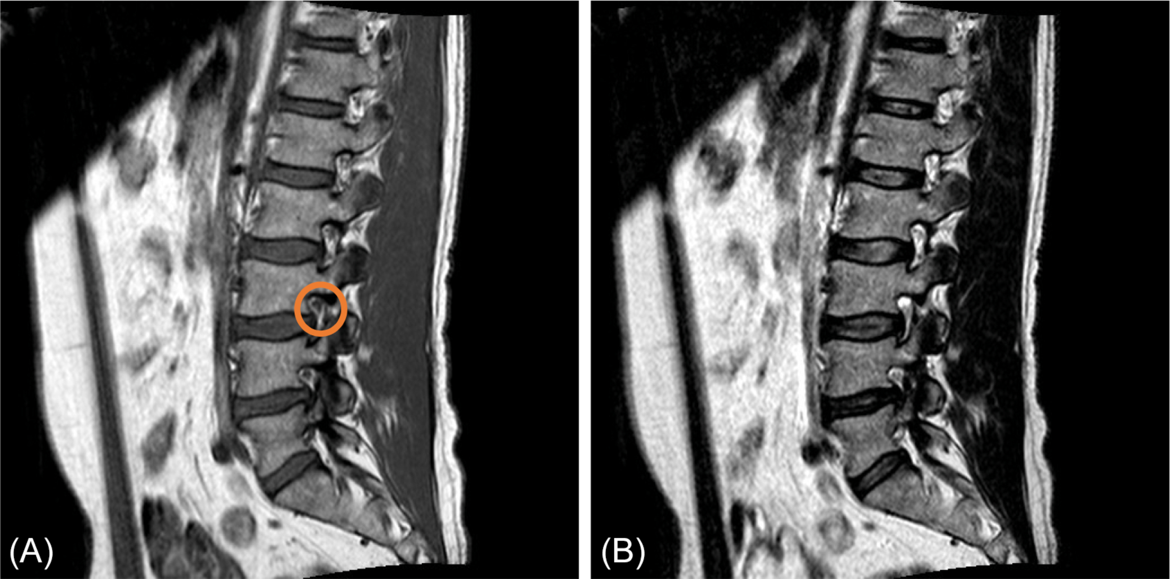
Lumbosacral Spine Mri Radiology Key
Change of address except Japan.
Does a lumbar spine mri show kidneys. It consists of twelve vertebrae which are separated by fibrocartilaginous intervertebral discs. 14700 Citicorp Drive Bldg. Magnetic resonance imaging MRI is a medical imaging technique used in radiology to form pictures of the anatomy and the physiological processes of the body in both health and disease. What is MRI and how does it work. Treatment of multiple myeloma focuses on decreasing the severity of symptoms with medications stem cell transplants. The journal has a broad International perspective and emphasises the advances occurring in Asia the Pacific Rim region Europe.
We are proud to present the latest paper of our ongoing series New Trends in Breast Imaging. This supports the lumbar spine in its main function as a weight bearing structure. This type of scan uses magnetic fields and radio waves in a non-invasive way to diagnose problems inside the body. Seek medical care for unexplained pain nausea vomiting weight loss vision problems or chronic tingling or numbness. Could monochromatic X-rays revolutionize breast imaging. For this test you lie on your side on a bed or exam table with your knees up near your chest.
Make no mistake about it these are rare herbal plant extracts that will help you effortlessly kickstart your type 2 diabetes reversal fills your. In rare cases the contrast dye used with MRI scans can cause severe allergic reactions or damage to your kidneys. The MRI is done to find tumors herniated discs or other soft-tissue disorders. Gadolinium is less likely to cause an allergic reaction than iodine contrast material. It produces detailed images of organs bones and soft tissue. As part of the bony thorax the thoracic vertebrae help protect the internal viscera such as the heart lungs and oesophagus.
It is made up of five distinct vertebrae which are the largest of the vertebral column. MRI does not use radiation and may require an injection of gadolinium contrast material. Multiple myeloma is a cancer of the plasma cells in the bone marrow. MRI is necessary to evaluate the spinal cord allowing differentiation of syrinx from for example a long-standing neoplasm. If the findings of the imaging studies are normal and the child is not tender to palpation and does not show evidence of a neurologic deficit the TL spine can be cleared. 3 Hagerstown MD 21742.
Lumbar and lower back pain can be treated by your spinal surgeon through fusion surgery the stabilization of both the back and front of the spine. It contains herbal extracts such as mistletoe and anacardium ocidentale. Middle back pain can be caused by strain from daily activities and poor posture a past or recent injury or muscle inflammation. The organs under the domain. Magnetic resonance imaging MRI of the spine uses radio waves a magnetic field and a computer. This may be limited to a portion of the spine often cervical but in severe cases extends inferiorly into the thoracic and even lumbar spine.
MRI is an abbreviation for magnetic resonance imaging. The lumbar spine is the third region of the vertebral column located in the lower back between the thoracic and sacral vertebral segments. There is no cure for multiple myeloma. This International journal Journal of Clinical Neuroscience publishes articles on clinical neurosurgery and neurology and the related neurosciences such as neuro-pathology neuro-radiology neuro-ophthalmology and neuro-physiology. The doctor first numbs an area in the lower part of the back near the spine. An MRI scan provides higher resolution images and better soft tissue contrast.
An MRI scan uses a powerful magnetic field and radio waves to take detailed 3-D pictures of your body. Otherwise conventional radiographs of the thoracic and lumbar spine should be obtained. Lumbar puncture spinal tap This test is used mainly to look for cancer cells in the cerebrospinal fluid CSF the liquid that surrounds the brain and spinal cord. Contains 40 tea bags To be taken 1 tea bag morning and evening Akum tea is a sure blend for the treatment of insulin dependent and non-insulin dependent diabetes. Having mid back pain is a common condition that can also feel like tightness or tension in the center of your back. The thoracic spine is the second segment of the vertebral column located between the cervical and lumbar vertebrae.
It can also demonstrate tonsillar herniation indicating Chiari malformation. An MRI will show the anatomy of the vertebrae as well as the disks spinal cord and the spaces between the vertebrae through which nerves pass. Matthias Dietzel MD MHBA Erlangen Germany In their groundbreaking work Michael Fishman PhD Boston University USA and Madan Rehani. The MRI is better than X-ray because in addition to the bones it can also show pictures of the nerves and discs. It creates clear detailed pictures of the spine and surrounding tissues. Read below for more information on why you may be having prolonged or sudden pain in the middle of your back.
The human body is 80 percent water so. The strong magnetic fields created during an MRI can cause heart pacemakers and other implants not to work as. The series is edited and supervised by our associated editor Dr. The MRI is the most commonly used test to evaluate the spine because it can show abnormal areas of the soft tissues around the spine. MRI magnetic resonance imaging and MR angiography Overview. Unlike X-rays or CT images are created by using a.
MRI creates images by distinguishing between the nuclear magnetic properties of various tissues a property that.

Magnetic Resonance Imaging Mri Of The Lumbosacral Spinal Cord A Download Scientific Diagram

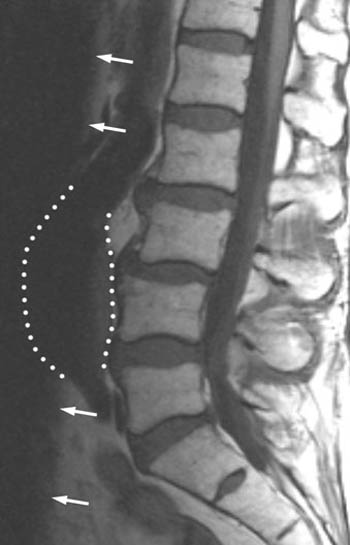
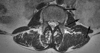
Lumbar Mri Shows Bilateral Renal Cysts Arrows On Axial T2 Weighted Download Scientific Diagram
Incidental Findings On Musculoskeletal Mr Radsource

Lumbar Mri Shows Uterine Myoma As Hypointense Lesion On Sagittal Download Scientific Diagram
Ajronline Org
Ajronline Org
Incidental Findings On Musculoskeletal Mr Radsource
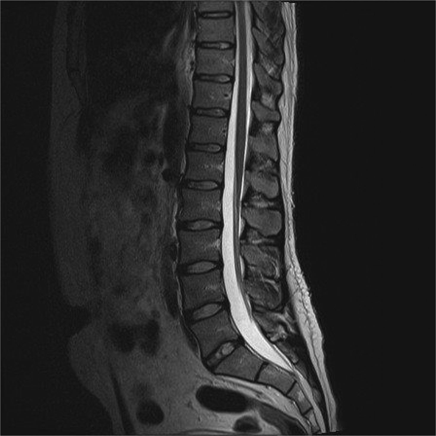
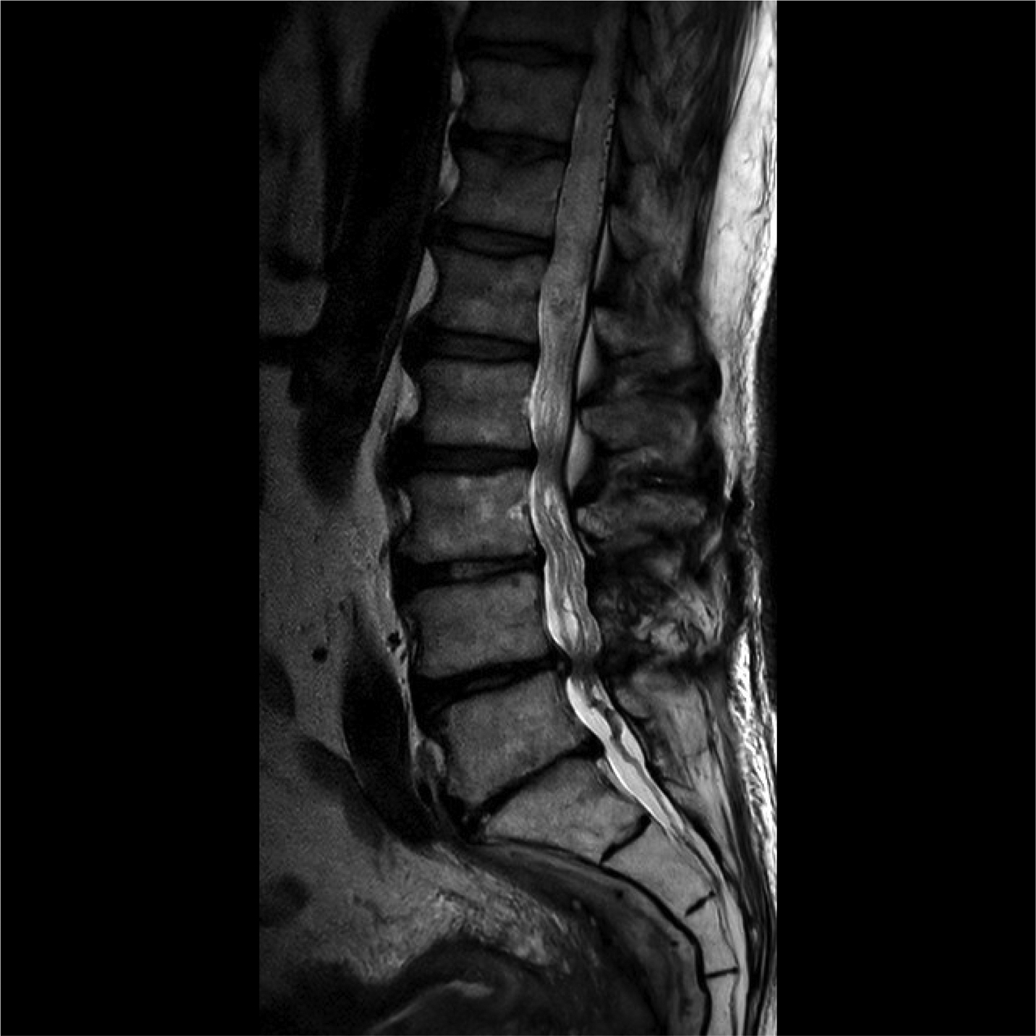
Lumbosacral Spine Mri Radiology Key

Mri Of Lumbar Spine Shows Hypointensity On T1wi A And Hyper Mix Download Scientific Diagram

Mri Of Lumbar Spine Shows Hypointensity On T1wi A And Hyper Mix Download Scientific Diagram

Do I Really Need An X Ray Or Mri For Lower Back Pain
Lumbosacral Spine Mri Radiology Key

Magnetic Resonance Imaging For Back Pain Mriplus

Color Mri Of The Lumbar Spine Low Back Showing A Normal Lumbar Spine And One With Disc Herniations Created By Medical Media Imag Disk Herniation Mri Anatomy
Posting Komentar untuk "Does A Lumbar Spine Mri Show Kidneys"