Bones Fused Across The Joint Space By Surgery
The different types of synovial joints are the ball-and-socket joint shoulder joint hinge joint knee pivot joint atlantoaxial joint between C1 and C2 vertebrae of the neck condyloid joint radiocarpal joint. The jaw articulates via a hinge joint between the quadrate and articular.
Pin On Cervical Spine

The other important concept to realize is that the spinal hardware is used to provide temporary fixation and.

Bones fused across the joint space by surgery. Heres what you need to know if you get infected. Hallux valgus usually called a bunion is a common deformity of the big toe that is predominantly seen in female patients. This is a debated topic. The opisthenar area dorsal is the corresponding area on the posterior part of the hand. For simple Lisfranc injuries a patient typically will do well with realignment of the bones. The Omicron variant has fueled a rise in Covid-19 cases across the United States.
These bones are the tibia the fibula and the talus. Elimination of motion across the disc space and reduction of loads on disc tissues theoretically result in. Should I have my injured foot realigned or fused. In advanced stages the deformity puts. Watch this video to see an animation of synovial joints in action. The separation of bones at these plates is a common occurrence in leg weakness or osteochondrosis particularly in young growing animals.
Most of the upper jaw bones premaxilla maxilla jugal quadratojugal and quadrate have been fused to the braincase while the lower jaw bones dentary splenial angular surangular and articular have been fused together into a unit called the mandible. Hardware placed for a fusion typically is not removed unless it becomes bothersome. Synovial joints are places where bones articulate with each other inside of a joint cavity. First of all the deformity is not pretty. CBCT enables to examine the joint space and the true position of the condyle within the fossa which is helpful in revealing likely dislocation of the joint disk. The hardware that is placed during surgery is sometimes removed 4-6 months after surgery.
By Dani Blum and Nicole Stock. Ankle arthritis is degeneration of the cartilage that covers the ends of the bones that form the ankle joint. A 53 yrs old man with the history of posttraumatic DDD at L3-L4 and L4-L5 underwent surgical repair. It is of particular concern to women whose feet no longer fit into shoes. The palm Volar which is the central region of the anterior part of the hand located superficially to the metacarpusThe skin in this area contains dermal papillae to increase friction such as are also present on the fingers and used for fingerprints. The big toe noticeably tilts toward the outside of the foot displacing the smaller toes.
Surgeon A performed an anterior exposure of the spine with the mobilization of the great vesselsSurgeon B performed anterior minimal discectomy and fusion at L3-L4 and L4-L5 using an anterior interbody technique. Near the ends of the bones are flattened areas of cartilage running at right angles to the bone called the epiphyseal plates which by increasing their thickness cause bones to grow in length and width. Moving the arthritic ankle tends to make the pain worse. In ankle fusion the ankle bones are fused into one bone. SurgiMend for plastic and reconstructive surgery muscle flap reinforcement hernia repair reinforcement of soft tissues repaired by sutures or suture anchors during tendon repair surgery including reinforcement of the rotator cuff patellar Achilles biceps quadriceps or other tendons and all other wound care indications for SurgiMend. Areas of the human hand include.
Surgery for asymptomatic closed spinal dysraphism in older individuals or clinical tethered cord syndrome without radiographic. Fusion of a diseased joint will eliminate pain by eliminating the motion between the painful joint such as severely diseased disks within the lumbar spine. 6 It usually takes 69 months for solid fusion to be seen radiographically. Additionally CBCT enables to quantify the roof of the glenoid fossa and assists in locating the soft tissue around the TMJ providing a practicable diagnosis and avoiding the. Lack of bridging bone or abnormal motion at fused segment after 12 months since fusion surgery or with.
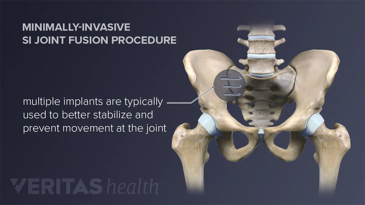
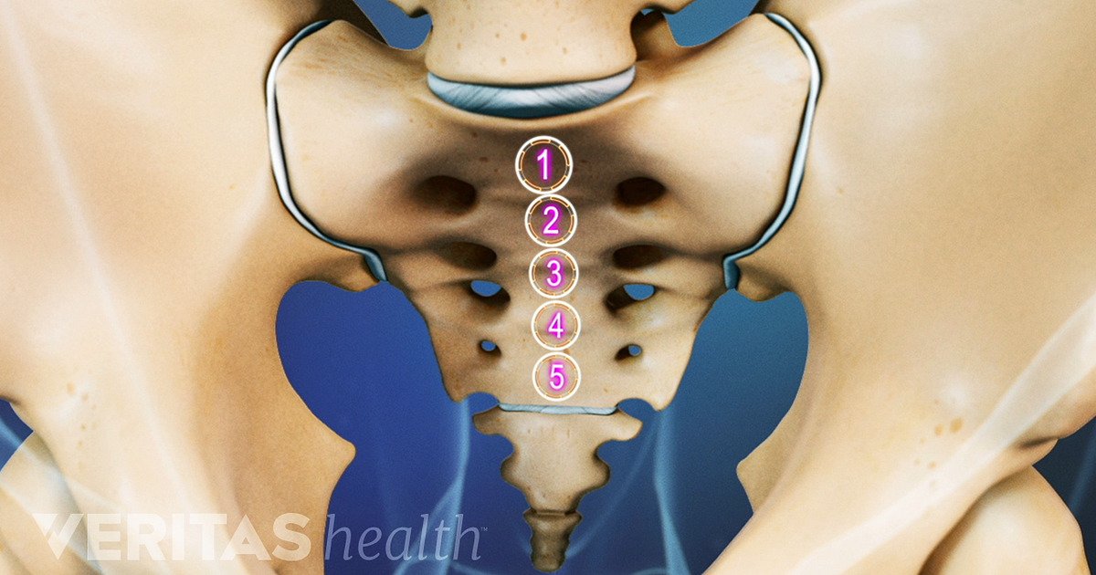
How Sacroiliac Joint Fusion Is Performed
Back Surgery L5 S1 Lumbar Discectomy And Spinal Fusion Stock Photo Royalty Free Image 7711632 Alamy Spinal Fusion Back Surgery Spinal
Nucleus Medical Media Spinal Fusion Surgery Spinal Fusion Spine Surgery

What To Know About Sacroiliac Joint Fusion
Fusing Together The Bones At The Shoulder Joint A Reasonable Alternative For Selective Desperate Situations Shoulder Elbow

Ankle Joint Replacement Surgery Is Common These Days The Process Include The Replacement Of Joint Replacement Ankle Replacement Ankle Strengthening Exercises

Subtalar Fusion Orthopaedic Associates Of Riverside
Laminectomy Spinal Fusion Fbss Failed Back Surgery Syndrome Yes This Is Me Spinal Fusion Back Surgery Spine Surgery

Acdf Surgery C3 6 Acdf Surgery Neck Surgery Spinal Fusion Surgery

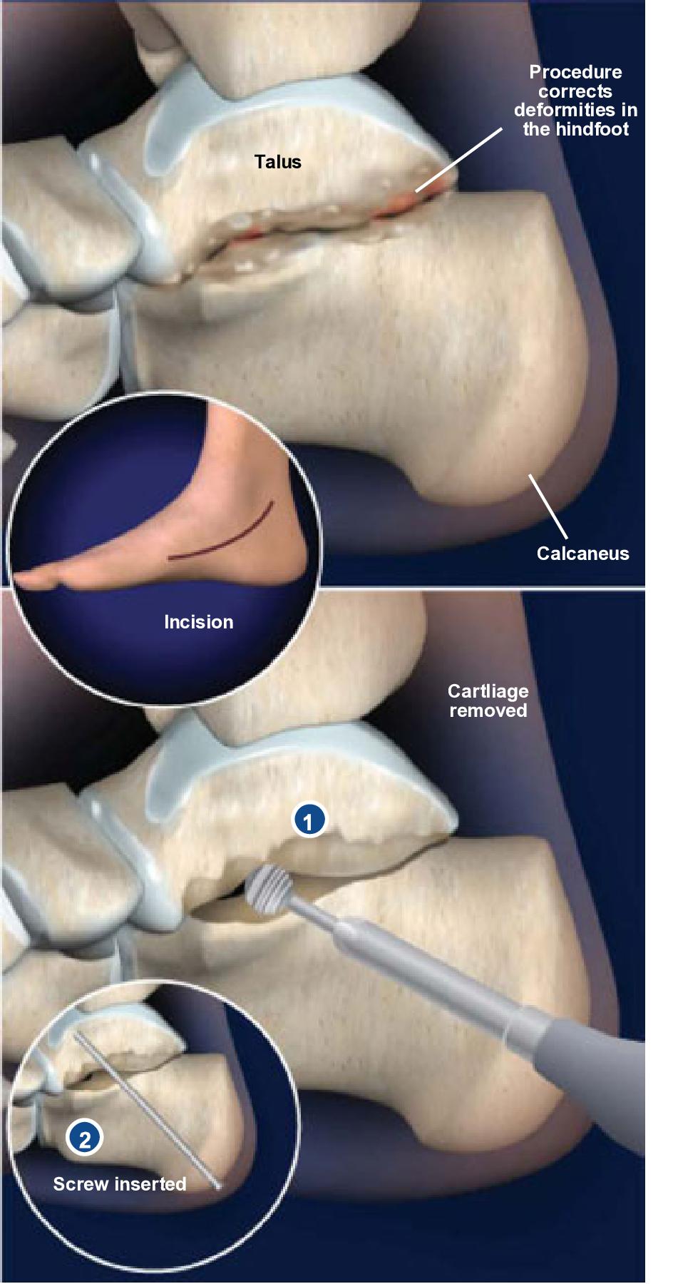
Triple Arthrodesis Surgery Foot And Ankle Specialist

Preparing For Lumbar Spinal Fusion Spinal Fusion Spinal Fusion Surgery Degenerative Disc Disease

Pin On Health

Pin On Auto Accident Injuries Issues

Fusing Together The Bones At The Shoulder Joint A Reasonable Alternative For Selective Desperate Situations Shoulder Elbow

Posting Komentar untuk "Bones Fused Across The Joint Space By Surgery"