What Does Ct Of Neck Show
These images show thin slices of your bones blood vessels and. During the exam contrast material is injected through a small catheter placed in a vein of the arm.

Pin On Ct Scans
What Does An Echocardiogram Show Patients With Chest Pain In patients with chest pain there are a number of different possible causes some of which can be assessed by echocardiography.

What does ct of neck show. Colon cancer however starts inside of the bowel. The US Preventive Services Task Force currently recommends low-dose CT without contrast along with appropriate patient counseling for patients with a history of smoking and an age range as detailed in the Task Force statement. This not enlarged according to the size criteria. CT manifestations of coronavirus disease-2019. Neck Pain Headaches. The palpable abnormality in the region of the right submandibular gland corresponds to a small-Volume lymph node measuring 1505cm.
Federal GovernmentRead our disclaimer for details. It outlines CT protocols for diagnostic imaging currently applied to ou CT scanners. For more details on the bones of the face please refer to the e-Anatomy module Face-CT-Scan. It can show large more advanced cancers or masses. If artery blockages are suspected the echocardiogram may show abnormalities in the walls of the heart supplied by those arteries. ClinicalTrialsgov is a resource provided by the US.
It can give. CT scan images provide more detailed information than plain X-rays do. An MRI uses radio waves and a strong magnet to create a series of detailed cross-sections of the soft tissues and bones. A computerized tomography CT scan combines a series of X-ray images taken from different angles and uses computer processing to create cross-sectional images or slices of the bones blood vessels and soft tissues inside your body. These tests can show damage and other issues in the bones and surrounding tissues in your neck. Slouching forward puts pressure between your shoulder blades and causes you to flatten your back muscles.
The EOCME is accredited by the Accreditation Council for. Whiplash occurs when the muscles in your neck suffer a strain because of a rapid movement backward and then forward. The bones of the face and neck were labeled using different colors to facilitate comprehension. To diagnose the cause of the pain your doctor may use imaging tests such as X-ray magnetic resonance imaging MRI or computed tomography CT. Binderow continues CT is very good for solid organ disease liver spleen kidneys. The Editors of Clinical Imaging in conjunction with the Elsevier Office of Continuing Medical Education are pleased to offer an AMA PRA Category 1 CME credit program for registered Clinical Imaging physician reviewers who complete manuscript reviews.
Computed tomography CT magnetic resonance imaging MRI CT angiography uses a CT scanner to produce detailed images of both blood vessels and tissues in various parts of the body. CT without contrast for screening. If you notice pain below the neck and around your tailbone after a long day at work you are likely not sitting up straight. A retrospective analysis of 73 cases by disease severity Liu et al. It also helps your doctor to evaluate your face sinuses and skull or to plan radiation therapy for brain cancer. By clicking on the link you will be leaving the official Royal Philips Healthcare Philips website.
AMA PRA Category 1 CME credit for Clinical Imaging reviewers. National Library of Medicine. The CT scan is an advanced form of X-ray machine that uses computers to create images of your inner neck. CT is notoriously unreliable for polyps or early stage tumors. It can show metastatic colon cancer that has spread to the liver. CT scans are very good at showing bone soft tissue and blood vessels Fig.
The diagnostic algorithm for lung cancer screening is evolving. 3 Follow-up of a solitary pulmonary. You have a swelling which you can feel palpable which is in the region of right side of the lower jaw right submandibular gland regionThis is 15 x 05 cm and this size is not. Neck CT images show focal or diffuse low-attenuation swelling of all or some of the soft tissues of the upper airway. LP Riccelli works closely with OHSU CT techs in the art of creating optimal images from current technology. This page is for Physicians Inside and outside this institution and CT Technologists.
While an MRI takes excellent pictures of soft tissue and blood vessels a CT scan shows bone much better so its often used to image the spine and skull. If available MRI is typically the first imaging option for viewing soft tissues because it does not have the risks associated with the additional radiation dose and myelography. For example focal involvement may be seen in the tongue Fig E5 particularly in association with angiotensin-converting enzyme inhibitors subcutaneous fat lips Fig 7 or soft palate and it may be unilateral 28. A computed tomography CT scan is often ordered if there is something abnormal on the chest X-ray. Listing a study does not mean it has been evaluated by the US. What does a CT show.
Soft-tissue tumors are defined as mesenchymal proliferations that occur in extraskeletal nonepithelial tissues of the body excluding the viscera meninges and lymphoreticular system 1 2CT has long been used to characterize the composition and anatomic location of soft-tissue masses 3-5 and has been known for several decades to be able to distinguish benign from. Poor posture puts pressure on your posterior muscles which has a negative impact on your neck. Computed tomography CT of the head uses special x-ray equipment to help assess head injuries severe headaches dizziness and other symptoms of aneurysm bleeding stroke and brain tumors. An MRI can be better at detecting abnormalities of the spinal cord bulging discs small disc herniations pinched nerves and other soft tissue problems. Any links to third-party websites that may appear on this site are provided only for your convenience and in no way represent any affiliation or endorsement of the information provided on those linked websites. European Journal of Radiology Vol126 108941.
See Computerized Tomography CT Scan MRI scan. A CT scan takes a cross-sectional and a more detailed image of the lung. Positron emission tomography-computed tomography PETCT with fluorine-18-fluorodeoxy-D-glucose FDG plays a major role today in the pre-therapeutic work-up and post-therapeutic monitoring of patients with head and neck tumoursFDG-PETCT is now routinely used in the head and neck for the delineation of the primary tumour detection of. MRI scans are better for imaging water-containing tissue. An MRI differs from a CAT scan also called a CT scan or a computed axial tomography scan because it does not use radiation. Before participating in a study talk to your health care provider and learn about the risks and potential benefits.
A Study of Debio 1143 Xevinapant in Combination With Platinum-Based Chemotherapy and Standard Fractionation Intensity-Modulated Radiotherapy in Participants With Locally Advanced Squamous Cell Carcinoma of the Head and Neck Suitable for Definitive Chemoradiotherapy - Full Text View. The sudden motion causes your necks tendons and ligaments to stretch and tear. The bone structures are rather more difficult to view on a weighted MRI T2 than on a CT-Scan. A radiologic technologist will capture high-resolution CT.

Pin On Ct Scans

Pin Op Ct Scans

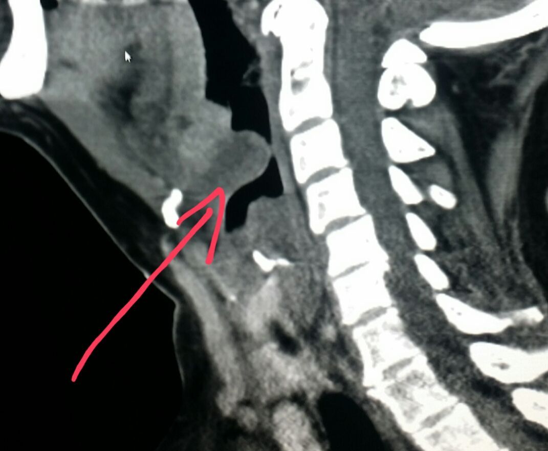
Thyroglossal Duct Cyst Reconstructed Ct Scan Of The Neck Demonstrates A Midline Cystic Lesion Red Arrow With A Slig Radiology Imaging Radiology Radiography
Pin On Ct Scans

Pin On Ct Scans

Pin En Coloana

Ct Neck Axial Anatomy Anatomy Anatomy Of The Neck Radiology Imaging

Ct Examination Of The Neck Are Done With The Iv Administration Of Contrast Media Artifacts Caused By Dental Work Often Obscure Ct Scan Radiology Head And Neck
Pin On Ct Scans

Pin On Ct Scans

Deep Spaces Of The Head And Neck Radiology Reference Article Radiopaedia Org Radiology Head And Neck Nursing School Notes

Pin On Ct Scans

Pin By Dr Abuaiad On Brain Head And Neck Pre And Post Radiology Head And Neck

Pin On Ct Scans
Posting Komentar untuk "What Does Ct Of Neck Show"